
 By
By 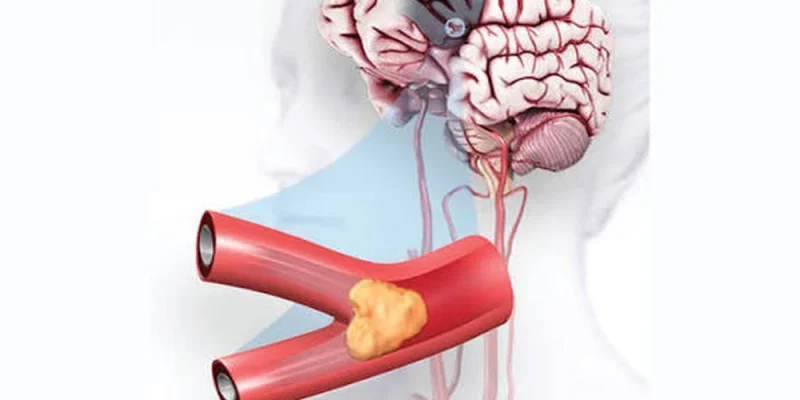
Stroke remains one of the leading causes of long-term disability worldwide, and outcomes hinge almost entirely on how fast it’s recognized and treated. But not all strokes are the same – and the specific type of stroke a patient is having determines which treatments can save brain tissue and which ones could cause catastrophic, irreversible harm. For nurses, understanding stroke subtypes isn’t academic trivia; it’s the difference between a life-saving intervention delivered in time and a preventable, fatal error.
Ischemic Stroke: Thrombotic and Embolic Subtypes
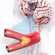
Roughly 87% of all strokes are ischemic, caused by a blockage that cuts off blood flow to part of the brain, starving neurons of oxygen and glucose within minutes. Ischemic strokes are further divided into thrombotic strokes, where a clot forms directly within a narrowed or diseased cerebral artery, and embolic strokes, where a clot forms elsewhere in the body – often in the heart due to atrial fibrillation – and then travels through the bloodstream to lodge in a brain vessel. Both mechanisms starve downstream brain tissue of oxygen, but their underlying causes, and sometimes the nuances of their treatment, differ meaningfully.
Hemorrhagic Stroke: Intracerebral and Subarachnoid Hemorrhage
Hemorrhagic strokes occur when a blood vessel in or around the brain ruptures, causing active bleeding into surrounding tissue. Intracerebral hemorrhage involves bleeding directly into brain parenchyma, often related to chronic, poorly controlled hypertension. Subarachnoid hemorrhage involves bleeding into the space surrounding the brain, frequently caused by a ruptured cerebral aneurysm and often presenting with a sudden, severe “thunderclap” headache. These strokes carry a notably high mortality rate and require an entirely different treatment approach than ischemic stroke – clot-busting medications used routinely for ischemic stroke would be catastrophic if given to a hemorrhagic stroke patient, which is why rapid imaging to distinguish the two is non-negotiable.
Transient Ischemic Attack (TIA): A Critical Warning Sign
A transient ischemic attack (TIA) is a temporary blockage that resolves on its own, often within minutes to an hour, without leaving permanent damage on imaging. TIAs produce classic stroke-like symptoms that fully resolve, but they are a critical warning sign rather than a reassuring “false alarm” – roughly one in three people who experience a TIA will go on to have a full, completed stroke, often within the following days. Treating a TIA as a near miss to be relieved about, rather than a medical emergency demanding urgent workup, is one of the more dangerous gaps in both patient and, occasionally, provider awareness.
Using the FAST Assessment Tool
The FAST acronym – Face drooping, Arm weakness, Speech difficulty, Time to call emergency services – remains the fastest, most reliable bedside tool for recognizing a possible stroke in progress. Nurses use it not only in dedicated emergency settings but during routine rounding and assessment, since strokes can occur in any inpatient unit, at any hour, in any patient. Rapid identification using FAST triggers formal stroke protocols that dramatically shorten the time from symptom onset to imaging and, ultimately, to treatment.
Time-Sensitive Treatment: tPA and Thrombectomy
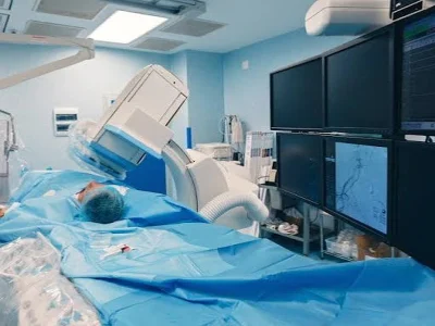
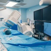
For ischemic stroke, treatment is a genuine race against the clock. Tissue plasminogen activator (tPA) can dissolve the offending clot if administered within a narrow window from symptom onset, and mechanical thrombectomy can physically retrieve larger clots in eligible patients, sometimes extending the treatment window further under specific imaging criteria. Hemorrhagic stroke management instead focuses on controlling ongoing bleeding, managing dangerously elevated intracranial pressure, and sometimes emergency surgical intervention to evacuate the hematoma. Every minute of delay in either pathway costs measurable, quantifiable brain function – a concept clinicians often summarize with the phrase “time is brain.”
Stroke Recovery and Rehabilitation
Recovery looks different depending on stroke type, brain location affected, and initial severity, but early, aggressive rehabilitation – physical, occupational, and speech therapy started as soon as medically appropriate – consistently improves long-term functional outcomes. Nurses play a central, ongoing role in secondary prevention too, reinforcing blood pressure control, anticoagulation or antiplatelet adherence, and lifestyle changes that meaningfully reduce the risk of a second, often more devastating, stroke. This full picture, from initial recognition through long-term recovery, is covered in the Strokes course, part of a broader lineup of accredited nursing courses online.
Risk Factors That Raise Stroke Likelihood
Stroke risk also compounds with certain patient populations more than others, which shapes both prevention counseling and clinical suspicion. Atrial fibrillation, uncontrolled hypertension, diabetes, and a prior TIA or stroke all meaningfully raise the likelihood of a future event, and nurses working in outpatient or chronic disease management settings play a real role in reinforcing the medications and lifestyle changes that reduce that risk over time. Recognizing which patients carry elevated baseline risk also sharpens clinical suspicion at the bedside, since a patient with known atrial fibrillation presenting with sudden confusion deserves a faster, more assertive stroke workup than the same symptom in a much lower-risk patient.
Clinical Scenario: Why Imaging Comes Before Treatment
Consider a scenario that illustrates why this distinction matters so much in practice: a patient arrives with sudden right-sided weakness and slurred speech. Family members aren’t sure exactly when symptoms started, and initial vitals show significantly elevated blood pressure. Before any thrombolytic can even be considered, a stat CT scan is mandatory to rule out hemorrhage, because giving tPA to a patient who is actually bleeding into their brain would be catastrophic rather than helpful. This single imaging step is why “time is brain” doesn’t mean skipping essential safety checks – it means moving through the correct sequence of assessment, imaging, and treatment as fast as possible, which is precisely why stroke protocols exist as standardized, rehearsed pathways rather than case-by-case improvisation.
Continuing Education for Stroke Recognition
Because stroke care intersects so closely with long-term blood pressure management, it pairs naturally with related coursework on how hypertension affects long-term organ health, available through the same nursing continuing education online catalog for nurses looking to build a more complete cardiovascular knowledge base. Whether you work in a dedicated stroke unit or simply want to sharpen your ability to recognize a stroke in progress on a general floor, understanding these subtypes at a mechanistic level, rather than as a list of memorized facts, builds the kind of clinical confidence that translates directly into faster, safer bedside decisions.






Comments